Lower Limb Bones
The Femur
The femur is the only bone in the thigh and the longest bone in the body. It acts as the site of origin and attachment of many muscles and ligaments, and can be divided into three parts; proximal, shaft and distal.
Proximal
The proximal aspect of the femur articulates with the acetabulum of the pelvis to form the hip joint.
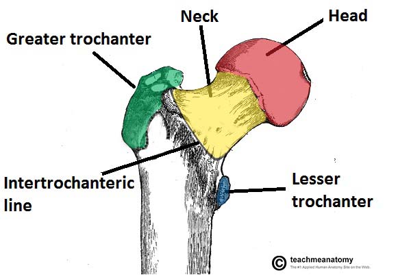
It consists of a head and neck, and two bony processes – the greater and lesser trochanters. There are also two bony ridges connecting the two trochanters; the intertrochanteric line anteriorly and the trochanteric crest posteriorly.
Head – articulates with the acetabulum of the pelvis to form the hip joint. It has a smooth surface, covered with articular cartilage (except for a small depression – the fovea – where ligamentum teres attaches).
Neck – connects the head of the femur with the shaft. It is cylindrical, projecting in a superior and medial direction. It is set at an angle of approximately 135 degrees to the shaft. This angle of projection allows for an increased range of movement at the hip joint.
Greater trochanter – the most lateral palpable projection of bone that originates from the anterior aspect, just lateral to the neck.
It is the site of attachment for many of the muscles in the gluteal region, such as gluteus medius, gluteus minimus and piriformis. The vastus lateralis originates from this site.
An avulsion fracture of the greater trochanter can occur as a result of forceful contraction of the gluteus medius.
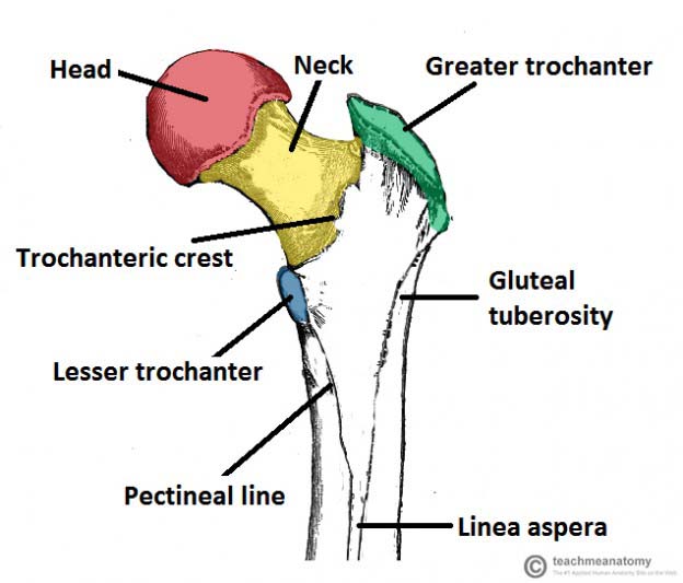
Lesser trochanter – smaller than the greater trochanter. It projects from the posteromedial side of the femur, just inferior to the neck-shaft junction.
- It is the site of attachment for iliopsoas (forceful contraction of which can cause an avulsion fracture of the lesser trochanter).
Intertrochanteric line – a ridge of bone that runs in an inferomedial direction on the anterior surface of the femur, spanning between the two trochanters. After it passes the lesser trochanter on the posterior surface, it is known as the pectineal line.
- It is the site of attachment for the iliofemoral ligament (the strongest ligament of the hip joint).
- It also serves as the anterior attachment of the hip joint capsule.
Intertrochanteric crest – like the intertrochanteric line, this is a ridge of bone that connects the two trochanters. It is located on the posterior surface of the femur. There is a rounded tubercle on its superior half called the quadrate tubercle; where quadratus femoris attaches.
The Shaft
The shaft of the femur descends in a slight medial direction. This brings the knees closer to the body’s centre of gravity, increasing stability. A cross section of the shaft in the middle is circular but flattened posteriorly at the proximal and distal aspects.
On the posterior surface of the femoral shaft, there are roughened ridges of bone, called the linea aspera (Latin for rough line). These split inferiorly to form the medial and lateral supracondylar lines. The flat popliteal surface lies between them.
Proximally, the medial border of the linea aspera becomes the pectineal line. The lateral border becomes the gluteal tuberosity, where the gluteus maximus attaches.
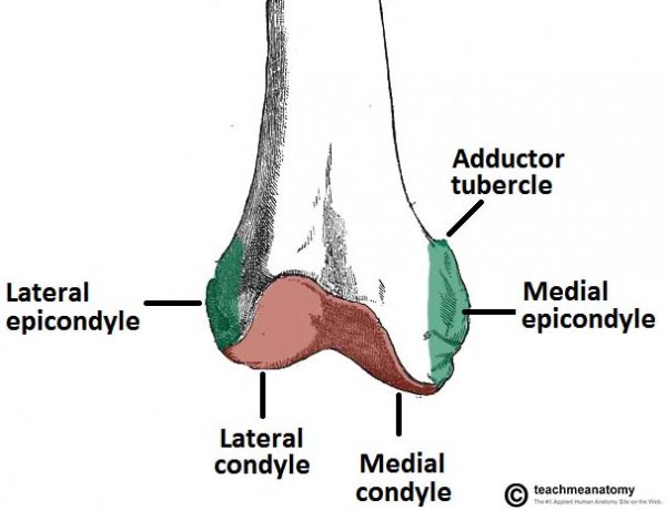
Distally, the linea aspera widens and forms the floor of the popliteal fossa, the medial and lateral borders form the medial and lateral supracondylar lines. The medial supracondylar line ends at the adductor tubercle, where the adductor magnus attaches.
Distal
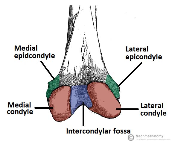
The distal end of the femur is characterised by the presence of the medial and lateral condyles, which articulate with the tibia and patella to form the knee joint.
Medial and lateral condyles – rounded areas at the end of the femur. The posterior and inferior surfaces articulate with the tibia and menisci of the knee, while the anterior surface articulates with the patella. The more prominent lateral condyle helps prevent the natural lateral movement of the patella; a flatter condyle is more likely to result in patellar dislocation.
Medial and lateral epicondyles – bony elevations on the non-articular areas of the condyles. The medial epicondyle is the larger.
- The medial and lateral collateral ligaments of the knee originate from their respective epicondyles.
Intercondylar fossa – a deep notch on the posterior surface of the femur, between the two condyles. It contains two facets for attachment of intracapsular knee ligaments; the anterior cruciate ligament (ACL) attaches to the medial aspect of the lateral condyle and the posterior cruciate ligament (PCL) to the lateral aspect of the medial condyle.
The Patella
The patella (knee-cap) is located at the front of the knee joint, within the patellofemoral groove of the femur. Its superior aspect is attached to the quadriceps tendon, and inferior aspect to the patellar ligament.
It is classified as a sesamoid type bone due to its position within the quadriceps tendon, and is the largest sesamoid bone in the body.
Bony Landmarks
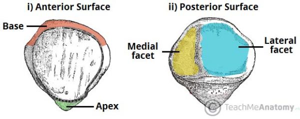
The patella has a triangular shape, with anterior and posterior surfaces. The apex of the patella is situated inferiorly, and is connected to the tibial tuberosity by the patella ligament. The base forms the superior aspect of the bone, and provides the attachment area for the quadriceps tendon.
The posterior surface of the patella articulates with the femur, and is marked by two facets:
- Medial facet – articulates with the medial condyle of the femur.
- Lateral facet – articulates with the lateral condyle of the femur.
Functions
The patella has two main functions:
- Leg extension – Enhances the leverage that the quadriceps tendon can exert on the femur, increasing the efficiency of the muscle.
- Protection – Protects the anterior aspect of the knee joint from physical trauma.
–
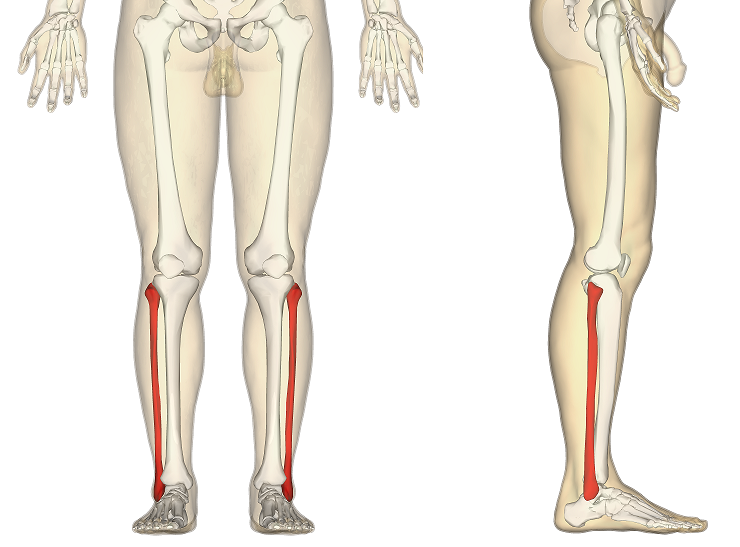
The Tibia
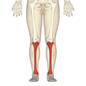
The tibia is the main bone of the lower leg, forming what is more commonly known as the shin.
It expands at its proximal and distal ends; articulating at the knee and ankle joints respectively. The tibia is the second largest bone in the body and it is a key weight-bearing structure.
Proximal
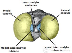
The proximal tibia is widened by the medial and lateral condyles, which aid in weight-bearing. The condyles form a flat surface, known as the tibial plateau. This structure articulates with the femoral condyles to form the key articulation of the knee joint.
Located between the condyles is a region called the intercondylar eminence – this projects upwards on either side as the medial and lateral intercondylar tubercles. This area is the main site of attachment for the ligaments and the menisci of the knee joint. The intercondylar tubercles of the tibia articulate with the intercondylar fossa of the femur.
Shaft
The shaft of the tibia is prism-shaped, with three borders and three surfaces; anterior, posterior and lateral. For brevity, only the anatomically and clinically important borders/surfaces are mentioned here.
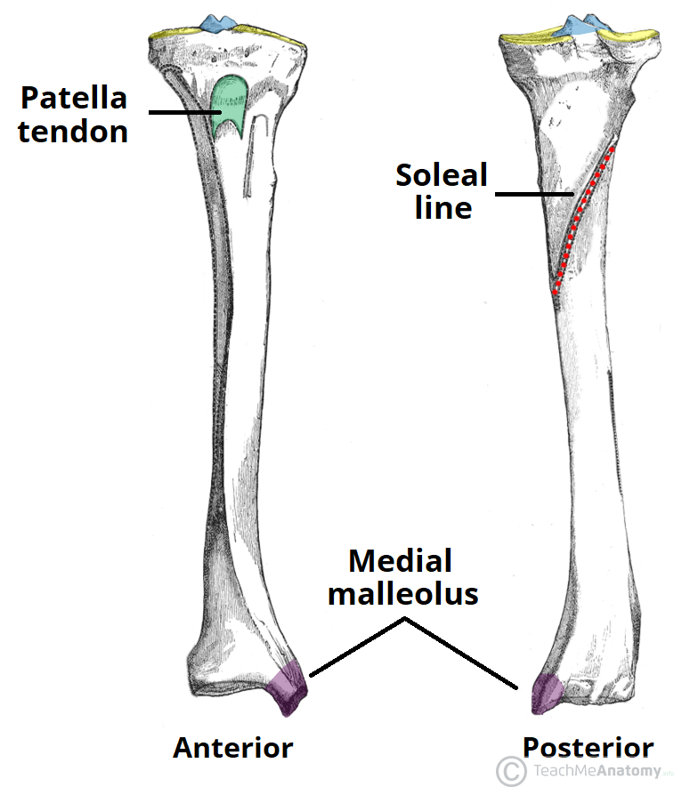
Anterior border – palpable subcutaneously down the anterior surface of the leg as the shin. The proximal aspect of the anterior border is marked by the tibial tuberosity; the attachment site for the patella ligament.
Posterior surface – marked by a ridge of bone known as soleal line. This line is the site of origin for part of the soleus muscle, and extends inferomedially, eventually blending with the medial border of the tibia. There is usually a nutrient artery proximal to the soleal line.
Lateral border – also known as the interosseous border. It gives attachment to the interosseous membrane that binds the tibia and the fibula together.
Distal
The distal end of the tibia widens to assist with weight-bearing.
The medial malleolus is a bony projection continuing inferiorly on the medial aspect of the tibia. It articulates with the tarsal bones to form part of the ankle joint. On the posterior surface of the tibia, there is a groove through which the tendon of tibialis posterior passes.
Laterally is the fibular notch, where the fibula is bound to the tibia – forming the tibiofibular joint.
Fibula
The fibula is a bone located within the lateral aspect of the leg. Its main function is to act as an attachment for muscles, and not as a weight-bearer.
It has three main articulations:
- Proximal tibiofibular joint – articulates with the lateral condyle of the tibia.
- Distal tibiofibular joint – articulates with the fibular notch of the tibia.
- Ankle joint – articulates with the talus bone of the foot.
Bony Landmarks
Proximal At the proximal end, the fibula has an enlarged head, which contains a facet for articulation with the lateral condyle of the tibia. On the posterior and lateral surface of the fibular neck, the common fibular nerve can be found.
Shaft The fibular shaft has three surfaces – anterior, lateral and posterior. The leg is split into three compartments, and each surface faces its respective compartment e.g anterior surface faces the anterior compartment of the leg.
Distal Distally, the lateral surface continues inferiorly, and is called the lateral malleolus. The lateral malleolus is more prominent than the medial malleolus, and can be palpated at the ankle on the lateral side of the leg.
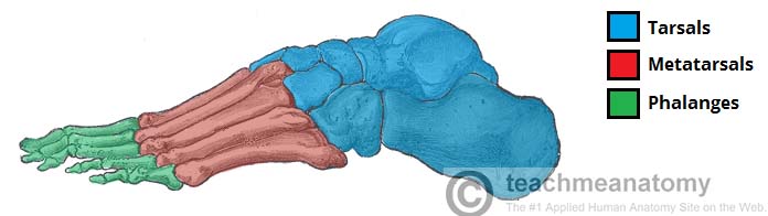
Bones of the Feet
They can be divided into three groups:
Tarsals – a set of seven irregularly shaped bones. They are situated proximally in the foot in the ankle area.
Metatarsals – connect the phalanges to the tarsals. There are five in number – one for each digit.
Phalanges – the bones of the toes. Each toe has three phalanges – proximal, intermediate and distal (except the big toe, which only has two phalanges).
The foot can also be divided up into three regions: 1. Hindfoot – talus and calcaneus 2. Midfoot – navicular, cuboid and cuneiforms 3. Forefoot – metatarsals and phalanges.
Tarsals
The tarsal bones of the foot are organised into three rows; proximal, intermediate and distal.
Proximal Group (Hindfoot)
The proximal tarsal bones are the talus and the calcaneus. These comprise the hindfoot, forming the bony framework around the proximal ankle and heel.
Talus
The talus is the most superior of the tarsal bones. It transmits the weight of the entire body to the foot. It has three articulations:
Superiorly – ankle joint – between the talus and the bones of the leg (the tibia and fibula).
Inferiorly – subtalar joint – between the talus and calcaneus.
Anteriorly – talonavicular joint – between the talus and the navicular.
The main function of the talus is to transmit forces from the tibia to the heel bone (known as the calcaneus). It is wider anteriorly compared to posteriorly which provides additional stability to the ankle.
Whilst numerous ligaments attach to the talus, no muscles originate from or insert onto it. This means there is a high risk of avascular necrosis as the vascular supply is dependent on fascial structures.
Calcaneus
The calcaneus is the largest tarsal bone and lies underneath the talus where it constitutes the heel. It has two articulations:
Superiorly – subtalar (talocalcaneal) joint – between the calcaneus and the talus.
Anteriorly – calcaneocuboid joint – between the calcaneus and the cuboid.
It protrudes posteriorly and takes the weight of the body as the heel hits the ground when walking. The posterior aspect of the calcaneus is marked by calcaneal tuberosity, to which the Achilles tendon attaches.
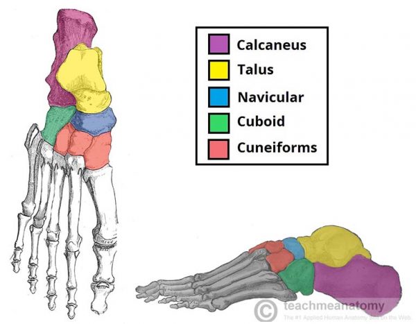
Intermediate Group (Midfoot)
The intermediate row of tarsal bones contains one bone, the navicular (given its name because it is shaped like a boat).
Positioned medially, it articulates with the talus posteriorly, all three cuneiform bones anteriorly, and the cuboid bone laterally. On the plantar surface of the navicular, there is a tuberosity for the attachment of part of the tibialis posterior tendon.
Distal Group (Midfoot)
In the distal row, there are four tarsal bones – the cuboid and the three cuneiforms. These bones articulate with the metatarsals of the foot
The cuboid is furthest lateral, lying anterior to the calcaneus and behind the fourth and fifth metatarsals. As its name suggests, it is cuboidal in shape. The inferior (plantar) surface of the cuboid is marked by a groove for the tendon of fibularis longus.
The three cuneiforms (lateral, intermediate (or middle) and medial) are wedge shaped bones. They articulate with the navicular posteriorly, and the metatarsals anteriorly. The shape of the bones helps form a transverse arch across the foot. They are also the attachment point for several muscles:
- Medial cuneiform – tibialis anterior, (part of) tibialis posterior and fibularis longus
- Lateral cuneiform – flexor hallucis brevis