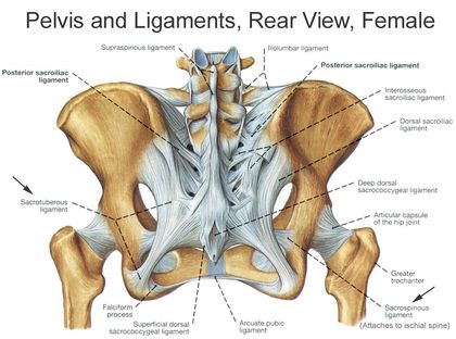
The major ligament that runs from the pelvis to the sacrum
The superior band extends over the sacroiliac joint and across the iliac crest to blend with the thoracolumbar fascia. The inferior band also passes over the anterior sacroiliac ligament to insert in the posterior region of the iliac fossa.
Sacroiliac
Anterior
Pre-auricular surface of the ilium to the third segment of the sacrum
It forms the anteroinferior component of the joint capsule.
Interosseous
It fills the gaps between the ilium and sacrum at the posterosuperior aspect of the joint
Deep to the posterior sacrioliac ligament
Posterior
This ligament covers the interosseous sacroiliac ligament as well as exiting dorsal rami of the sacral nerves.
It forms the communication between the posterior superior iliac spine, as well as part of the iliac crest to the lateral and intermediate sacral crests.
Sacrospinous
Margins of the coccyx and sacrum to the spine of the ischium.
Sacrotuberous
Attachments to the posterior sacroiliac ligaments, lower transverse tubercles of the sacrum, the posterior superior iliac spine, the proximal part of the coccyx and the lower lateral margins of the sacrum.
Pubic Ligament
Superior
Extends laterally from one pubic tubercle to the other.
The stability of the pubic symphysis is reinforced by the superior and inferior (arcuate) pubic ligaments.
Inferior
Extends laterally from one pubic tubercle to the other.
The stability of the pubic symphysis is reinforced by the superior and inferior (arcuate) pubic ligaments.
Inguinal Ligament
Lacunar Ligament
Spans the space between the inguinal and pectineal ligaments
The crescentic structure also forms the medial border of the femoral canal.
Pectineal Ligament
Continuation of the lacunar ligament along the pectineal line of the pubic bone.
It forms the posterior border of the femoral canal.
Inguinal Ligament
Anterior superior iliac spine to the ipsilateral pubic tubercle.
This fibrous structure goes on to form the floor of the inguinal canal.
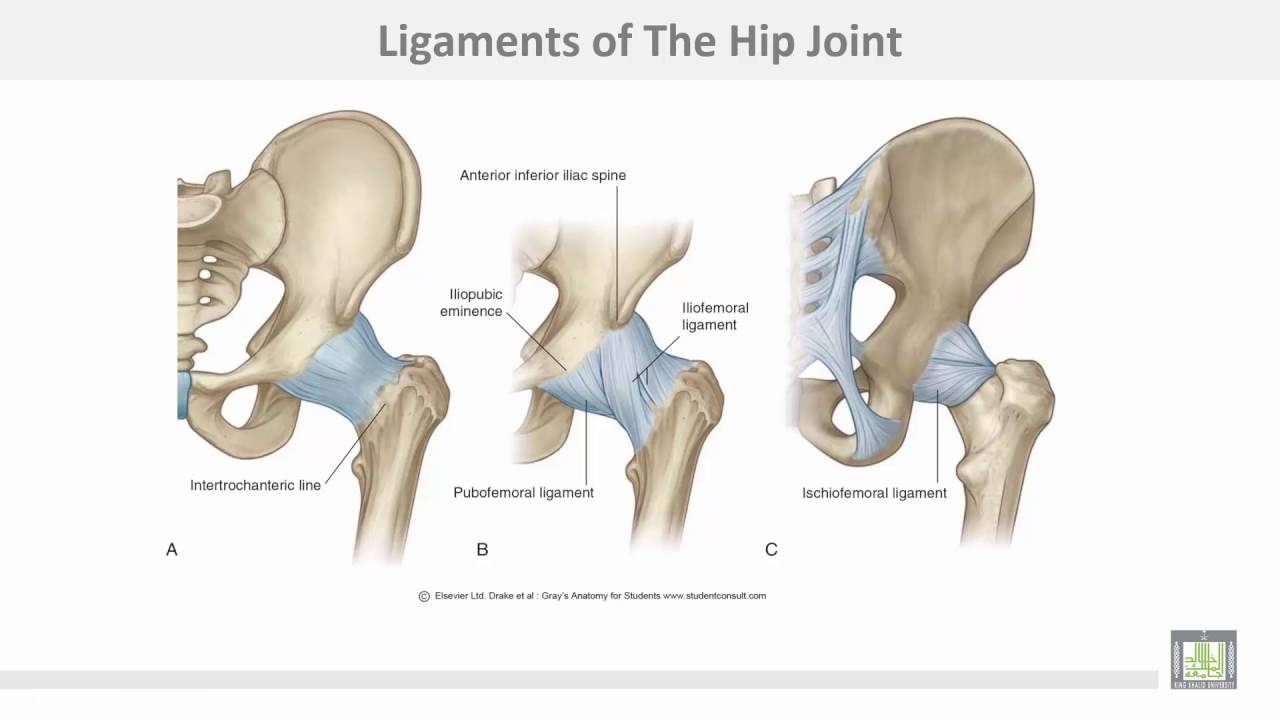
Hip Joint
The Hip
Ligament Group
Ligament
Description
Notes
Transverse acetabular ligament
Completes the labrum along the inferior border of the acetabulum.
In its course the ligament crosses the acetabular notch, converting it into the acetabular foramen.
Ligamentum Teres Caput Femoris
Its apex attaches to the fovea of the head of the femur, while its base inserts around the acetabular notch (at the center of the acetabulum).
Plays a role in anchoring the head of the femur in the acetabulum.
Pubofemoral
Proximally: obturator crest & membrane, superior pubic ramus and the iliopubic eminence. Distally: iliofemoral ligament (and occasionally the neck of the femur).
Iliofemoral (Y Ligament Of Bigelow)
Extends from its basal attachment at the intertrochanteric line to its apex between the rim of the acetabulum and the anterior inferior iliac spine.
Ischiofemoral
Cebntral Superior
Extends from the greater trochanter of the femur to the ischium (posteroinferior to the acetabulum)
Found at the posterior aspect of the joint.
Medial
Circumscribe the neck of the femur posteriorly.
Found at the posterior aspect of the joint.
Lateral
Circumscribe the neck of the femur posteriorly.
Found at the posterior aspect of the joint.
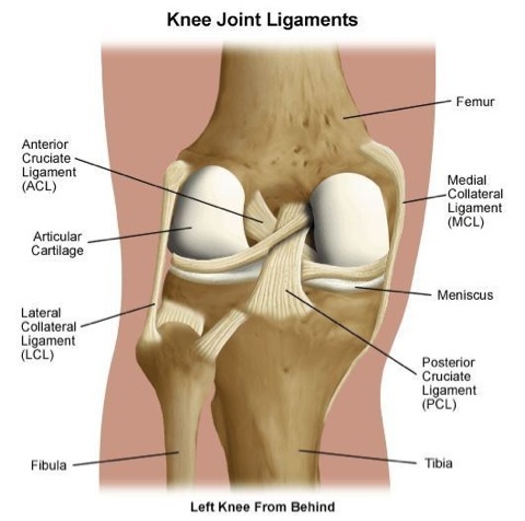
Knee and Tibiofibular Joints
The KneeThe Leg
Ligament Group
Ligament
Description
Notes
Cruciate Ligaments
Anterior
Medial tibial eminence and inserts in the posteromedially on the lateral femoral condyle.
Can be further subdivided (on microscopic dissection) into the posterolateral, anteromedial and intermediate bundles.
Posterior
Posterior intercondylar area of the tibia and inserts in the lateral part of the medial femoral condyle and as far as the anterior region of the intercondylar notch.
Meniscofemoral Ligaments
Anterior
Posterior horn of the lateral meniscus and joins with the posterior cruciate ligament to insert in the lateral aspect of the medial femoral condyle.
Travels posterior to the posterier cruciate ligament
Posterior
Posterior horn of the lateral meniscus and joins with the posterior cruciate ligament to insert in the lateral aspect of the medial femoral condyle.
Travels anterior to the posterier cruciate ligament
Transverse Ligament of the Knee
Communicate horizontally between the anterior aspects of the horns of the medial and lateral menisci.
Superior tibiofibular ligaments
Posterior
Solitary band that travels superomedially from the posterior aspect of the fibular head to the lateral condyle of the tibia.
Adds further stability to the joint
Anterior
Three bands that also take a superomedial course from the anterior part of the head of the fibula to the lateral condyle of the tibia.
Adds further stability to the joint
Collateral Ligaments
Medial (Tibial)
Originates just inferior to the adductor tubercle of the medial epicondyle of the femur to the medial epicondyle of the tibia.
Counteracts valgus (medially directed) forces
Lateral (Fibular)
Lateral femoral condyle to the head of the fibula.
Counteracts varus (laterally directed) forces
Patellar Ligament
Connects the patella to the tibia.
The ligament is a common point for clinicians to test the reflexes at the knee.
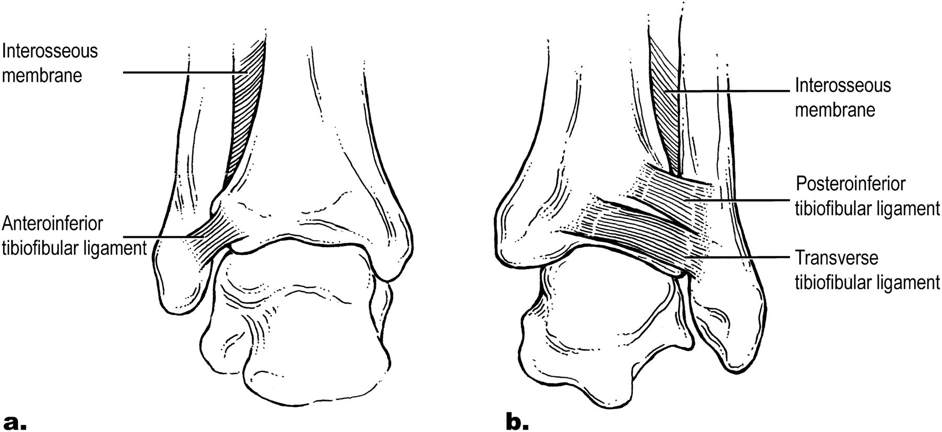
Ankle Joint and Foot
The Ankle
THIS NEEDS TO BE FIXED!!!!!!!!!!!!!!!!!!!!!!!!!!!!!!!!!!!!!!!!!!!!!!!!!!!!!!!!!!!!!!!!!!!!!!!!!!!!!!!!!!!!!!!!!!!!!!!!!!!!!!
Ligament Group
Ligament
Description
Notes
Cruciate Ligaments
Anterior
Medial tibial eminence and inserts in the posteromedially on the lateral femoral condyle.
Can be further subdivided (on microscopic dissection) into the posterolateral, anteromedial and intermediate bundles.
Posterior
Posterior intercondylar area of the tibia and inserts in the lateral part of the medial femoral condyle and as far as the anterior region of the intercondylar notch.
Meniscofemoral Ligaments
Anterior
Posterior horn of the lateral meniscus and joins with the posterior cruciate ligament to insert in the lateral aspect of the medial femoral condyle.
Travels posterior to the posterier cruciate ligament
Posterior
Posterior horn of the lateral meniscus and joins with the posterior cruciate ligament to insert in the lateral aspect of the medial femoral condyle.
Travels anterior to the posterier cruciate ligament
Transverse Ligament of the Knee
Communicate horizontally between the anterior aspects of the horns of the medial and lateral menisci.
Superior tibiofibular ligaments
Posterior
Solitary band that travels superomedially from the posterior aspect of the fibular head to the lateral condyle of the tibia.
Adds further stability to the joint
Anterior
Three bands that also take a superomedial course from the anterior part of the head of the fibula to the lateral condyle of the tibia.
Adds further stability to the joint
Collateral Ligaments
Medial (Tibial)
Originates just inferior to the adductor tubercle of the medial epicondyle of the femur to the medial epicondyle of the tibia.
Counteracts valgus (medially directed) forces
Lateral (Fibular)
Lateral femoral condyle to the head of the fibula.
Counteracts varus (laterally directed) forces
Patellar Ligament
Connects the patella to the tibia.
The ligament is a common point for clinicians to test the reflexes at the knee.