Lower Limb Muscles
The Fascia Lata
Fascia is a sheet or band of fibrous tissue lying deep to the skin. It lines, invests, and separates structures within the body. There are three main types of fascia:
- Superficial fascia – blends with the reticular layer beneath the dermis.
- Deep fascia – envelopes muscles, bones, and neurovascular structures.
- Visceral fascia – provides membranous investments that suspend organs within their cavities.
The fascia lata is a deep fascial investment of the musculature of the thigh, and is analogous to a strong, extensible, and elasticated stocking. It begins proximally around the iliac crest and inguinal ligament, and ends distal to the bony prominences of the tibia. It is continuous with what is renamed the deep fascia of the leg (also known as the crural fascia).
The depth of the fascia lata varies considerably across the thigh. It is thickest along the superolateral aspect of the thigh, where it arises from the fascial condensations of gluteus maximus and medius. It is also thick around the knee where the fascia receives reinforcing fibres from tendons of the quadriceps muscles. The fascial investment is thinnest where it covers the adductor muscles of the medial thigh.
The deepest aspect of the fascia lata gives rise to three intermuscular septa that attach centrally to the femur. The septa divide the thigh musculature into three compartments; anterior, medial, and lateral. The lateral intermuscular septum is the strongest of the three due to reinforcement from the iliotibial tract (see later), whereas the other two septa are proportionately weaker.
An ovoid hiatus known as the saphenous opening is present in the fascia lata just inferior to the inguinal ligament. The opening serves as an entry point for efferent lymphatic vessels and the great saphenous vein, draining into superficial inguinal lymph nodes and the femoral vein respectively.
| Item | Movement | Compartmentalisation | Muscular sheath |
|---|---|---|---|
| Iliotibial Tract (ITT) | Acts as an extensor, abductor and lateral rotator of the hip, with an additional role in providing lateral stabilisation to the knee joint. | TYhe deepest aspect of ITT extends centrally to form the lateral intermuscular septum of the thigh and attaches to the femur. | Forms a sheath around the tensor fascia lata muscle. |
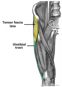
The tensor fascia lata is a gluteal muscle that acts as a flexor, abductor, and internal rotator of the hip. Its name, however, is derived from its additional role in tensing the fascia lata. It is innervated by the superior gluteal nerve, like gluteus medius and minimus, but is located more anterolaterally than the other gluteal muscles.
The muscle originates from the iliac crest, and descends inferiorly to the superolateral thigh. At the junction of the middle and upper thirds of the thigh, it inserts into the anterior aspect of the iliotibial tract. When stimulated, the tensor fasciae lata tautens the iliotibial band and braces the knee, especially when the opposite foot is lifted.
The property of TFL tightening the fascia lata is analogous to hoisting an elastic stocking up the thigh. When the fascia lata is pulled taut, it forces muscles in the anterior and posterior compartments closer to towards the femur. Contraction within each compartment centralises muscle weight and limits outward expansion, which in turn reduces the overall force required for movement at the hip joint.
An additional property of tensing the fascia lata is that it makes muscle contraction more efficient in compressing deep veins, which ensures adequate venous return to the heart from the lower limbs.
Proximal The fascia lata arises from multiple superior attachments around the pelvis and hip region:
- Posterior – sacrum and coccyx.
- Lateral – iliac crest.
- Anterior – inguinal ligament, superior pubic rami.
- Medial – inferior ischiopubic rami, ischial tuberosity, sacrotuberous ligament.
The fascia lata is also continuous with other regions of deep and superficial fascia at its superior aspect. The deep iliac fascia descends from the thoracic region at the diaphragm, covers the entire iliacus and psoas regions, and blends with the fascia lata superiorly.
The superficial fascia of the inferior abdominal wall (Scarpa’s fascia) and perineal region both blend with the fascia lata just below the inguinal ligament.
Lateral
The lateral thickening of fascia lata forms the iliotibial tract and receives tendon insertions superiorly from gluteus maximus and tensor fascia lata. The widened band of fibres descends the lateral thigh and attaches to the lateral tibial condyle on the anterolateral (Gerdy) tubercle.
Inferior
The fascia lata ends at the knee joint where it then becomes the deep fascia of the leg (the crural fascia). Attachments are made at bony prominences around the knee including the femoral and tibial condyles, patella, head of fibula and the tibial tuberosity.
Central
The deep aspect of fascia lata produces three intermuscular septa which attach centrally to the femur. The lateral septum joins to the lateral lip of the linea aspera and the medial and anterior septa attach to the medial lip. These attachments then continue along the whole length of the femur to include the supracondylar lines.
Thigh Anterior Compartment Muscles
The gluteal region is an anatomical area located posteriorly to the pelvic girdle, at the proximal end of the femur. The muscles in this region move the lower limb at the hip joint.
The muscles of the gluteal region can be broadly divided into two groups:
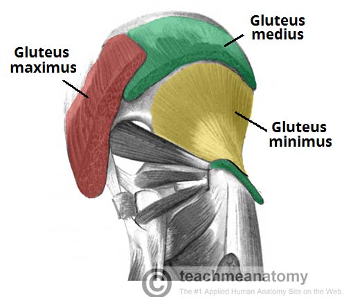
Superficial abductors and extenders – group of large muscles that abduct and extend the femur. Includes the gluteus maximus, gluteus medius, gluteus minimus and tensor fascia lata.
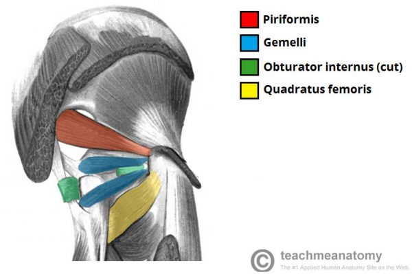
Deep lateral rotators – group of smaller muscles that mainly act to laterally rotate the femur. Includes the quadratus femoris, piriformis, gemellus superior, gemellus inferior and obturator internus.
The arterial supply to these muscles is mostly via the superior and inferior gluteal arteries – branches of the internal iliac artery. Venous drainage follows the arterial supply.
| Group | Muscles | Attachments | Actions | Innervation | Notes |
|---|---|---|---|---|---|
| Superficial Muscles | Gluteus Maximus | Originates from the gluteal (posterior) surface of the ilium, sacrum and coccyx. It slopes across the buttock at a 45 degree angle, then inserts into the iliotibial tract and the gluteal tuberosity of the femur. | It is the main extensor of the thigh, and assists with lateral rotation. However, it is only used when force is required, such as running or climbing. | Inferior gluteal nerve. | The gluteus maximus is the largest of the gluteal muscles. It is also the most superficial, producing the shape of the buttocks. |
| Gluteus Medius | Originates from the gluteal surface of the ilium and inserts into the lateral surface of the greater trochanter. | Abducts and medially rotates the lower limb. During locomotion, it secures the pelvis, preventing pelvic drop of the opposite limb. | Superior gluteal nerve. | The gluteus medius muscle is fan-shaped and lies between to the gluteus maximus and the minimus. It is similar in shape and function to the gluteus minimus. | |
| Gluteus Minimus | Originates from the ilium and converges to form a tendon, inserting to the anterior side of the greater trochanter. | Abducts and medially rotates the lower limb. During locomotion, it secures the pelvis, preventing pelvic drop of the opposite limb. | Superior gluteal nerve. | The gluteus minimus is the deepest and smallest of the superficial gluteal muscles. It is similar is shape and function to the gluteus medius. | |
| Tensor Fascia Lata | Originates from the anterior iliac crest, attaching to the anterior superior iliac spine (ASIS). It inserts into the iliotibial tract, which itself attaches to the lateral condyle of the tibia. | Assists the gluteus medius and minimus in abduction and medial rotation of the lower limb. It also plays a supportive role in the gait cycle. | Superior gluteal nerve. | Tensor fasciae lata is a small superficial muscle which lies towards the anterior edge of the iliac crest. It functions to tighten the fascia lata, and so abducts and medially rotates the lower limb. | |
| Deep Muscles | Piriformis | Originates from the anterior surface of the sacrum. It then travels infero-laterally, through the greater sciatic foramen, to insert into the greater trochanter of the femur. | Lateral rotation and abduction. | Nerve to piriformis. | The piriformis muscle is a key landmark in the gluteal region. It is the most superior of the deep muscles. |
| Obturator Internus | Originates from the pubis and ischium at the obturator foramen. It travels through the lesser sciatic foramen, and attaches to the greater trochanter of the femur. | Lateral rotation and abduction. | Nerve to obturator internus. | The obturator internus forms the lateral walls of the pelvic cavity. In some texts, the obturator internus and the gemelli muscles are considered as one muscle – the triceps coxae. | |
| The Gemelli – Superior and Inferior | The superior gemellus muscle originates from the ischial spine, the inferior from the ischial tuberosity. They both attach to the greater trochanter of the femur. | Lateral rotation and abduction. | The superior gemellus muscle is innervated by the nerve to obturator internus, the inferior gemellus is innervated by the nerve to quadratus femoris. | The gemelli are two narrow and triangular muscles. They are separated by the obturator internus tendon. | |
| Quadratus Femoris | It originates from the lateral side of the ischial tuberosity, and attaches to the quadrate tuberosity on the intertrochanteric crest. | Lateral rotation. | Nerve to quadratus femoris. | The quadratus femoris is a flat, square-shaped muscle. It is the most inferior of the deep gluteal muscles, located below the gemelli and obturator internus. |
Thigh Anterior Compartment Muscles
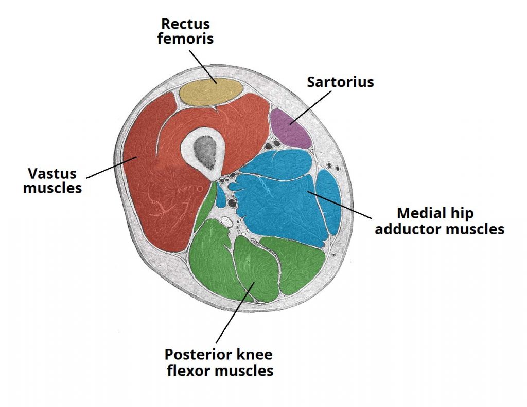
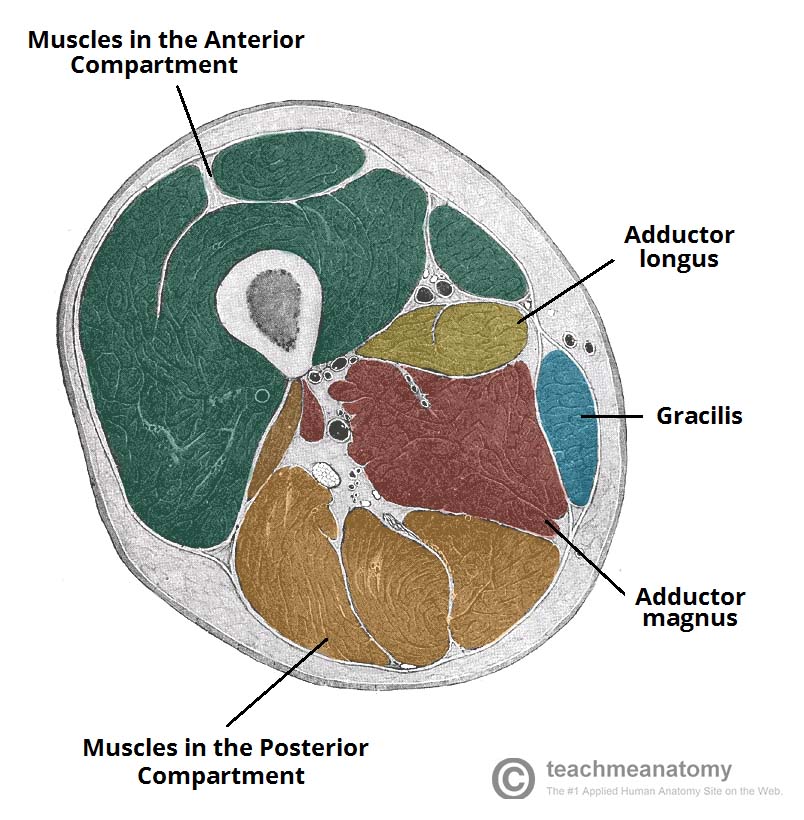
The musculature of the thigh can be split into three sections; anterior, medial and posterior. Each compartment has a distinct innervation and function. The muscles in the anterior compartment of the thigh are innervated by the femoral nerve (L2-L4), and as a general rule, act to extend the leg at the knee joint.
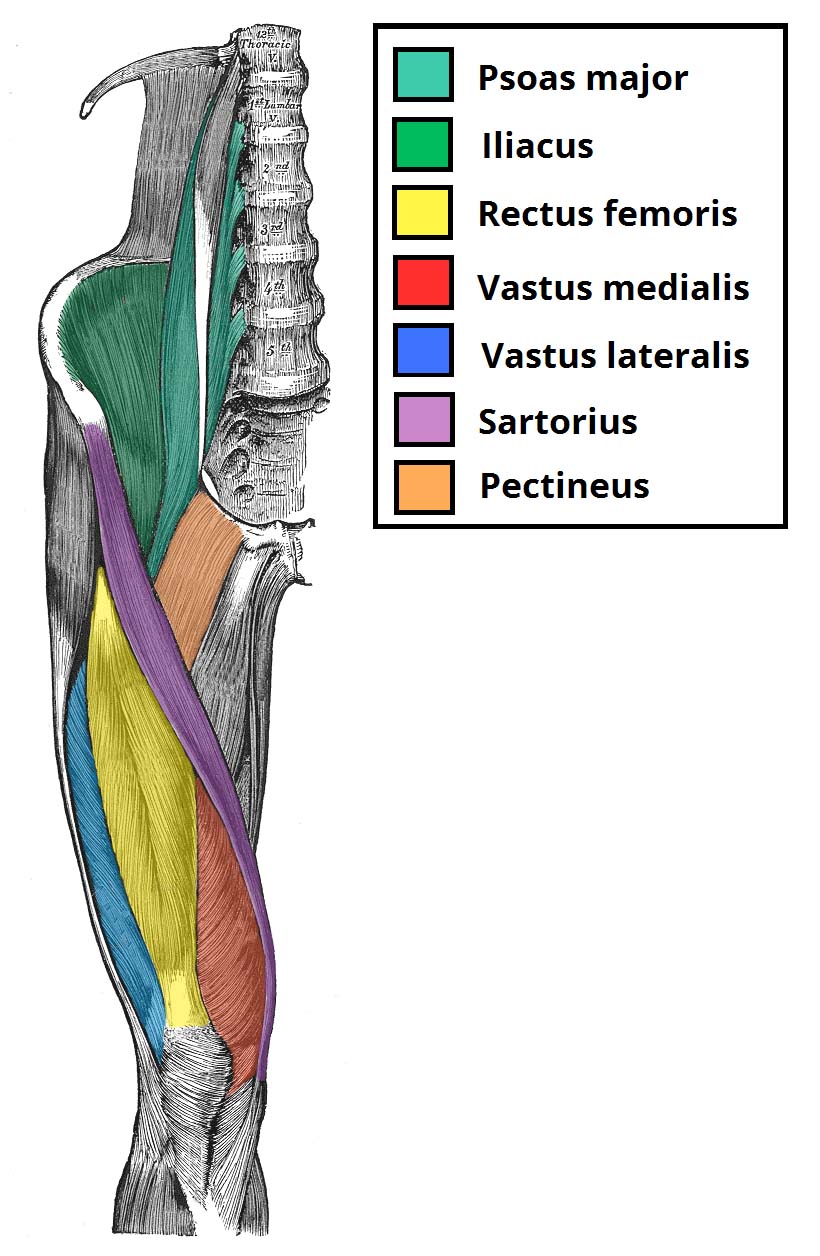
There are three major muscles in the anterior thigh – the pectineus, sartorius and quadriceps femoris. In addition to these, the end of the iliopsoas muscle passes into the anterior compartment.
| Muscle Group | Muscles | Attachments | Actions | Innervation | Notes |
|---|---|---|---|---|---|
| Iliopsoas | Psoas Major | The psoas major originates from the lumbar vertebrae. Inserts onto the lesser trochanter of the femur. | Flexes the thigh at the hip joint. | The psoas major is innervated by anterior rami of L1-3. | The iliopsoas is actually two muscles, the psoas major and the iliacus. They originate in different areas, but come together to form a tendon, hence why they are commonly referred to as one muscle. |
| Iliacus | Originates from the iliac fossa of the pelvis. Inserts onto the lesser trochanter of the femur. | Flexes the thigh at the hip joint. | The Iliacus is innervated by the femoral nerve. | The iliopsoas is actually two muscles, the psoas major and the iliacus. They originate in different areas, but come together to form a tendon, hence why they are commonly referred to as one muscle. | |
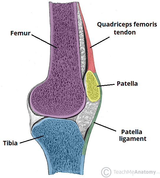
| Quadriceps Femoris | Vastus Lateralis | Proximal attachment: Originates from the greater trochanter and the lateral lip of linea aspera. | Extends the knee joint and stabilises the patella. | Femoral nerve. | The quadriceps femoris consists of four individual muscles; three vastus muscles and the rectus femoris. They form the main bulk of the thigh, and collectively are one of the most powerful muscles in the body. |
| Vastus Intermedius | Proximal attachment: Anterior and lateral surfaces of the femoral shaft. | Extends the knee joint and stabilises the patella. | Femoral nerve. | The quadriceps femoris consists of four individual muscles; three vastus muscles and the rectus femoris. They form the main bulk of the thigh, and collectively are one of the most powerful muscles in the body. | |
| Vastus Medialis | Proximal attachment: The intertrochanteric line and medial lip of the linea aspera. | Extends the knee joint and stabilises the patella, particularly due to its horizontal fibres at the distal end. | Femoral nerve. | The quadriceps femoris consists of four individual muscles; three vastus muscles and the rectus femoris. They form the main bulk of the thigh, and collectively are one of the most powerful muscles in the body. | |
| Rectus Femoris | Originates from the ilium, just superior to the acetabulum. It runs straight down the leg and attaches to the patella by the quadriceps femoris tendon. | The only muscle of the quadriceps to cross both the hip and knee joints. It flexes the thigh at the hip joint, and extends at the knee joint. | Femoral nerve. | The quadriceps femoris consists of four individual muscles; three vastus muscles and the rectus femoris. They form the main bulk of the thigh, and collectively are one of the most powerful muscles in the body. | |
| Sartorius | Originates from the anterior superior iliac spine, and attaches to the superior, medial surface of the tibia. | At the hip joint, it is a flexor, abductor and lateral rotator. At the knee joint, it is also a flexor. | Femoral nerve. | The sartorius is the longest muscle in the body. It is long and thin, running across the thigh in a inferomedial direction. The sartorius is positioned more superficially than the other muscles in the leg. | |
| Pectineus | It originates from the pectineal line on the anterior surface of the pelvis, and attaches to the pectineal line on the posterior side of the femur, just inferior to the lesser trochanter. | Adduction and flexion at the hip joint. | Femoral nerve. May also receive a branch from the obturator nerve. | The pectineus muscle is a flat muscle that forms the base of the femoral triangle. It has a dual innervation, and thus can be considered a transitional muscle between the anterior thigh and medial thigh compartments. |
Thigh Posterior Compartment Muscles
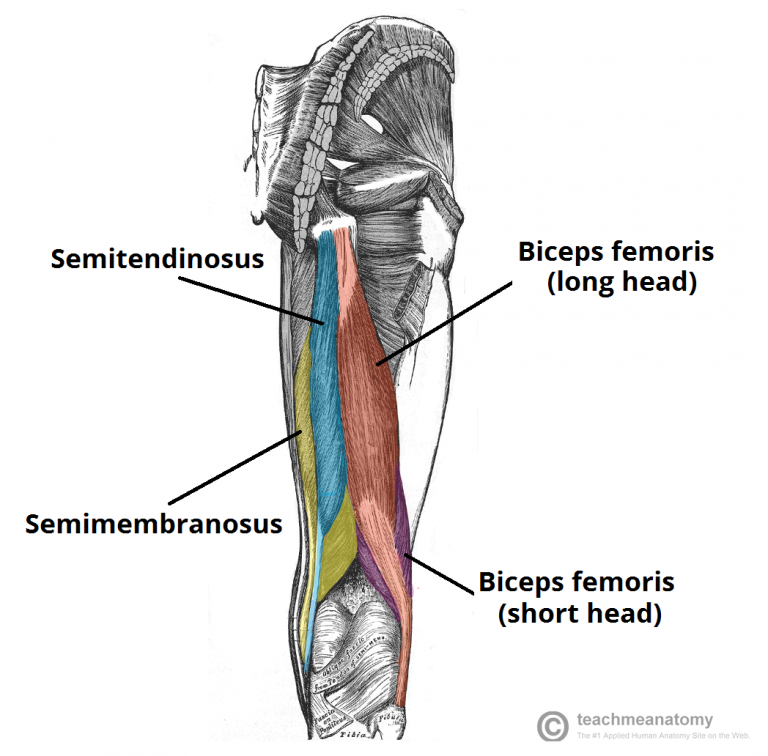
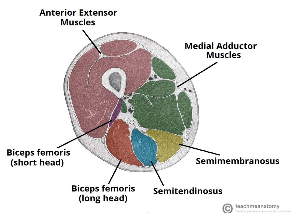
The muscles in the posterior compartment of the thigh are collectively known as the hamstrings. They consist of the biceps femoris, semitendinosus and semimembranosus, which form prominent tendons medially and laterally at the back of the knee.
As group, these muscles act to extend at the hip, and flex at the knee. They are innervated by the sciatic nerve (L4-S3).
| Muscles | Attachments | Actions | Innervation | Notes |
|---|---|---|---|---|
| Biceps Femoris | The long head originates from the ischial tuberosity of the pelvis. The short head originates from the linea aspera on posterior surface of the femur. Together, the heads form a tendon, which inserts into the head of the fibula. | Main action is flexion at the knee. It also extends the thigh at the hip, and laterally rotates at the hip and knee. | Long head innervated by the tibial part of the sciatic nerve, whereas the short head is innervated by the common fibular part of the sciatic nerve. | Like the biceps brachii in the arm, the biceps femoris muscle has two heads – a long head and a short head. It is the most lateral of the muscles in the posterior thigh |
| Semitendinosus | It originates from the ischial tuberosity of the pelvis, and attaches to the medial surface of the tibia. | Flexion of the leg at the knee joint. Extension of thigh at the hip. Medially rotates the thigh at the hip joint and the leg at the knee joint. | Tibial part of the sciatic nerve. | The semitendinosus is a largely tendinous muscle. It lies medially to the biceps femoris, and covers the majority of the semimembranosus. |
| Semimembranosus | It originates from the ischial tuberosity, but does so more superiorly than the semitendinosus and biceps femoris. It attaches to the medial tibial condyle. | Flexion of the leg at the knee joint. Extension of thigh at the hip. Medially rotates the thigh at the hip joint and the leg at the knee joint. | Tibial part of the sciatic nerve. | The semimembranosus muscle is flattened and broad. It is located underneath the semitendinosus. |
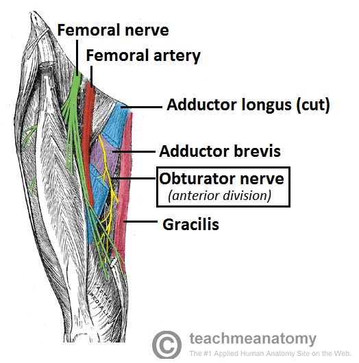
Thigh Medial Compartment Muscles
The muscles in the medial compartment of the thigh are collectively known as the hip adductors. There are five muscles in this group; gracilis, obturator externus, adductor brevis, adductor longus and adductor magnus.
All the medial thigh muscles are innervated by the obturator nerve, which arises from the lumbar plexus. Arterial supply is via the obturator artery.
| Muscles | Part | Attachments | Actions | Innervation | Notes |
|---|---|---|---|---|---|
| Adductor Magnus | Adductor | Originates from the inferior rami of the pubis and the rami of ischium, attaching to the linea aspera of the femur. | Adducts and flexes thigh | Adductor part is innervated by the obturator nerve (L2-L4) | The adductor magnus is the largest muscle in the medial compartment. It lies posteriorly to the other muscles. Functionally, the muscle can be divided into two parts; the adductor part, and the hamstring part. |
| Hamstring | Originates from the ischial tuberosity and attaches to the adductor tubercle and medial supracondylar line of the femur. | Adducts and extends thigh | Hamstring part is innervated by the tibial component of the sciatic nerve (L4-S3). | The adductor magnus is the largest muscle in the medial compartment. It lies posteriorly to the other muscles. Functionally, the muscle can be divided into two parts; the adductor part, and the hamstring part. | |
| Adductor Longus | Originates from the pubis, and expands into a fan shape, attaching broadly to the linea aspera of the femur | Adduction of the thigh. | Obturator nerve (L2-L4). | The adductor longus is a large, flat muscle. It partially covers the adductor brevis and magnus. The muscle forms the medial border of the femoral triangle. | |
| Adductor Brevis | Originates from the body of pubis and inferior pubic rami. It attaches to the linea aspera on the posterior surface of the femur, proximal to the adductor longus. | Adduction of the thigh. | Obturator nerve (L2-L4). | The adductor brevis is a short muscle, lying underneath the adductor longus. It lies in between the anterior and posterior divisions of the obturator nerve. | |
| Obturator Externus | It originates from the membrane of the obturator foramen, and adjacent bone. It passes under the neck of femur, attaching to the posterior aspect of the greater trochanter. | Adduction and lateral rotation of the thigh. | Obturator nerve (L2-L4). | This is one of the smaller muscles of the medial thigh, and it is located most superiorly. | |
| Gracilis | It originates from the inferior rami of the pubis, and the body of the pubis. It attaches to the medial surface of the tibia, between the tendons of the sartorius (anteriorly) and the semitendinosus (posteriorly). | Adduction of the thigh at the hip, and flexion of the leg at the knee. | Obturator nerve (L2-L4). | The gracilis is the most superficial and medial of the muscles in this compartment. It crosses at both the hip and knee joints. It is sometimes transplanted into the hand or forearm to replace a damaged muscle. |
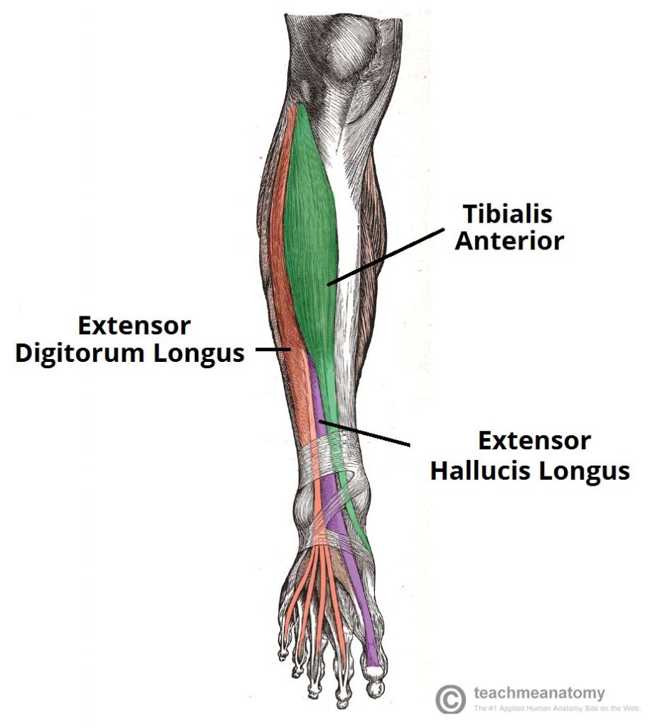
Lower Leg Anterior Compartment Muscles
There are four muscles in the anterior compartment of the leg; tibialis anterior, extensor digitorum longus, extensor hallucis longus and fibularis tertius.
Collectively, they act to dorsiflex and invert the foot at the ankle joint. The extensor digitorum longus and extensor hallucis longus also extend the toes. The muscles in this compartment are innervated by the deep fibular nerve (L4-S1), and blood is supplied via the anterior tibial artery.
| Muscle | Attachments | Actions | Innervation |
|---|---|---|---|
| Tibialis Anterior | Originates from the lateral surface of the tibia, attaches to the medial cuneiform and the base of metatarsal 1. | Dorsiflexion and inversion of the foot. | Deep fibular nerve. |
| Extensor Digitorum Longus | Originates from the lateral condyle of the tibia and the medial surface of the fibula. | Extension of the lateral four toes, and dorsiflexion of the foot. | Deep fibular nerve. |
| Extensor Hallucis Longus | Originates from the medial surface of the fibular shaft and attaches to the base of the distal phalanx of the great toe. | Extension of the great toe and dorsiflexion of the foot. | Deep fibular nerve. |
| Fibularis Tertius | Originates with the extensor digitorum longus from the medial surface of the fibula and attaches to metatarsal 5. | Eversion and dorsiflexion of the foot. | Deep fibular nerve. |
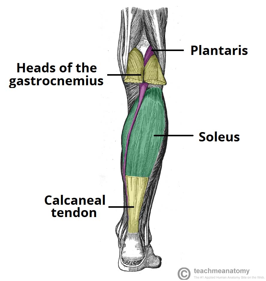
Lower Leg Posterior Compartment Muscles
The posterior compartment of the leg contains seven muscles, organised into two layers – superficial and deep. The two layers are separated by a band of fascia.
The posterior leg is the largest of the three compartments. Collectively, the muscles in this area plantarflex and invert the foot. They are innervated by the tibial nerve, a terminal branch of the sciatic nerve.
| Muscle | Layer | Attachments | Actions | Innervation |
|---|---|---|---|---|
| Gastrocnemius | Superficial | The lateral head originates from the lateral femoral condyle, and medial head from the medial femoral condyle. They attach to the calcaneus. | It plantarflexes at the ankle joint, and because it crosses the knee, it is a flexor there. | Tibial nerve. |
| Plantaris | Superficial | Originates from the lateral supracondylar line of the femur and attaches to the calcaneus. | It plantarflexes at the ankle joint, and because it crosses the knee, it is a flexor there. | Tibial nerve. |
| Soleus | Superficial | Originates from the soleal line of the tibia and proximal fibular area and attaches to the calcaneus. | Plantarflexes the foot at the ankle joint. | Tibial Nerve. |
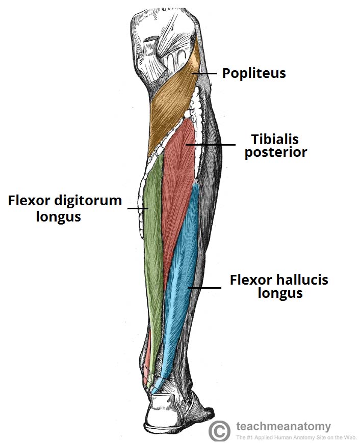
| Popliteus | Deep | Originates from the lateral condyle of the femur and the posterior horn of the lateral meniscus and insterts above the origin of the soleus muscle. | Laterally rotates the femur on the tibia – ‘unlocking’ the knee joint so that flexion can occur. | Tibial nerve. |
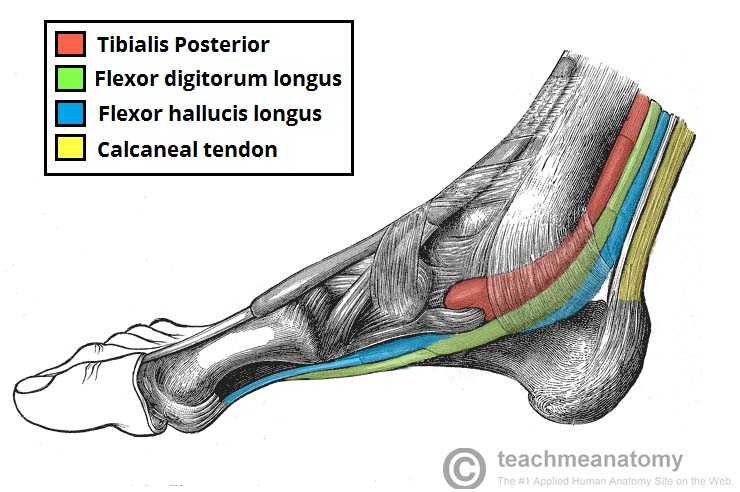
| Tibialis Posterior | Deep | Originates from the interosseous membrane between the tibia and fibula, and posterior surfaces of the two bones and attaches to the plantar surfaces of the medial tarsal bones. | Inverts and plantarflexes the foot, maintains the medial arch of the foot. | Tibial nerve. |
| Flexor Digitorum Longus | Deep | Originates from the medial surface of the tibia, attaches to the plantar surfaces of the lateral four digits. | Flexes the lateral four toes. | Tibial nerve. |
| Flexor Hallucis Longus | Deep | Originates from the posterior surface of the fibula, attaches to the plantar surface of the phalanx of the great toe. | Flexes the great toe. | Tibial nerve. |
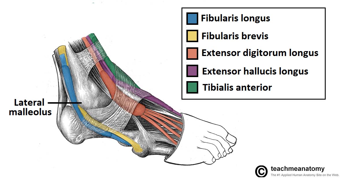
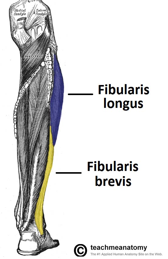
Lower Leg Lateral Compartment Muscles
There are two muscles in the lateral compartment of the leg; the fibularis longus and brevis (also known as peroneal longus and brevis).
The common function of the muscles is eversion – turning the sole of the foot outwards. They are both innervated by the superficial fibular nerve.
| Muscle | Attachments | Actions | Innervation |
|---|---|---|---|
| Fibularis Longus | The fibularis longus originates from the superior and lateral surface of the fibula and the lateral tibial condyle. It attaches to the medial cuneiform and base of metatarsal 1. | Eversion and plantarflexion of the foot. Also supports the lateral and transverse arches of the foot. | Superficial fibular (peroneal) nerve, L4-S1. |
| Fibularis Brevis | Originates from the inferolateral surface of the fibular shaft and attcachesto a tubercle on metatarsal 5. | Eversion of the foot. | Superficial fibular (peroneal) nerve, L4-S1. |
Foot Muscles
The muscles acting on the foot can be divided into two distinct groups; extrinsic and intrinsic muscles.
The extrinsic muscles arise from the anterior, posterior and lateral compartments of the leg. They are mainly responsible for actions such as eversion, inversion, plantarflexion and dorsiflexion of the foot.
The intrinsic muscles are located within the foot and are responsible for the fine motor actions of the foot, for example movement of individual digits.
| Compartment | Muscles | Layer | Attachments | Actions | Innervation | Notes |
|---|---|---|---|---|---|---|
| Dorsal Aspect | Extensor Digitorum Brevis | Originates from the calcaneus, the interosseous talocalcaneal ligament and the inferior extensor retinaculum. It attaches to the long extensor tendons of the four lateral digits. | Aids the extensor digitorum longus in extending the lateral four toes at the metatarsophalangeal and interphalangeal joints. | Deep fibular nerve. | The extensor digitorum brevis muscle lies deep to the tendon of the extensor digitorum longus. | |
| Extensor Hallucis Brevis | Originates from the calcaneus, the interosseous talocalcaneal ligament and the inferior extensor retinaculum. It attaches to the base of the proximal phalanx of the great toe. | Aids the extensor hallucis longus in extending the great toe at the metatarsophalangeal joint. | Deep fibular nerve. | Some texts consider the extensor hallucis brevis to be merely the medial part of the extensor digitorum brevis | ||
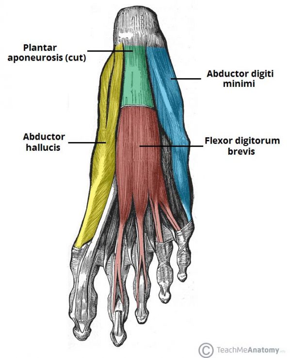
| Plantar Aspect | Abductor Hallucis | First Layer | Originates from the medial tubercle of the calcaneus, the flexor retinaculum and the plantar aponeurosis. It attaches to the medial base of the proximal phalanx of the great toe. | Abducts and flexes the great toe. | Medial plantar nerve. | The abductor hallucis muscle is located on the medial side of the sole, where it contributes to a small soft tissue bulge. |
| Flexor Digitorum Brevis | First Layer | Originates from the medial tubercle of the calcaneus and the plantar aponeurosis. It attaches to the middle phalanges of the lateral four digits. | Flexes the lateral four digits at the proximal interphalangeal joints. | Medial plantar nerve. | The flexor digitorum brevis muscle is located laterally to the abductor hallucis. It sits in the centre of the sole, sandwiched between the plantar aponeurosis and the tendons of flexor digitorum longus. | |
| Abductor Digiti Minimi | First Layer | Originates from the medial and lateral tubercles of the calcaneus and the plantar aponeurosis. It attaches to the lateral base of the proximal phalanx of the 5th digit. | Abducts and flexes the 5th digit. | Lateral plantar nerve. | The abductor digiti minimi muscle is located on the lateral side of the foot. It is homologous with the abductor digiti minimi of the hand. | |
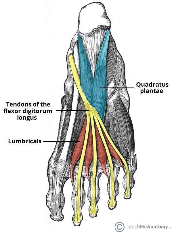
| Quadratus Plantae | Second Layer | Originates from the medial and lateral plantar surface of the calcaneus. It attaches to the tendons of flexor digitorum longus. | Assists flexor digitorum longus in flexing the lateral four digits. | Lateral plantar nerve. | The quadratus plantae muscle is located superior to the flexor digitorum longus tendons. It is separated from the first layer of muscles by the lateral plantar vessels and nerve. | |
| Lumbricals | Second Layer | Originates from the tendons of flexor digitorum longus. Attaches to the extensor hoods of the lateral four digits. | Flexes at the metatarsophalangeal joints, while extending the interphalangeal joints. | The most medial lumbrical is innervated by the medial plantar nerve. The remaining three are innervated by the lateral plantar nerve. | There are four lumbrical muscles in the foot. They are each located medial to their respective tendon of the flexor digitorum longus. | |
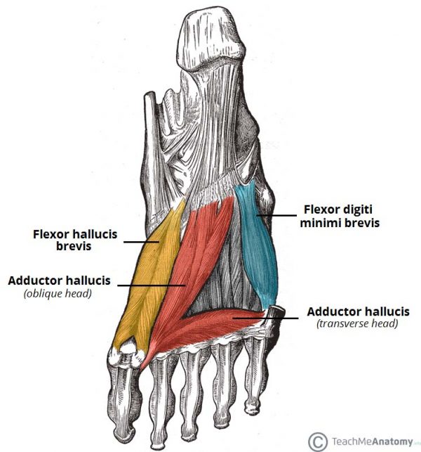
| Flexor Hallucis Brevis | Third Layer | Originates from the plantar surfaces of the cuboid and lateral cuneiforms, and from the tendon of the posterior tibialis tendon. Attaches to the base of the proximal phalanx of the great toe. | Flexes the proximal phalanx of the great toe at the metatarsophalangeal joint. | Medial plantar nerve. | The flexor hallucis brevis muscle is located on the medial side of the foot. It originates from two places on the sole of the foot. | |
| Adductor Hallucis | Third Layer | The oblique head originates from the bases of the 2nd, 3rd and 4th metatarsals. The transverse head originates from the plantar ligaments of the metatarsophalangeal joints. Both heads attach to the lateral base of the proximal phalanx of the great toe. | Adduct the great toe. Assists in forming the transverse arch of the foot. | Deep branch of lateral plantar nerve. | The adductor hallucis muscle is located laterally to the flexor hallucis brevis. It consists of an oblique and transverse head. | |
| Flexor Digiti Minimi Brevis | Third Layer | Originates from the base of the fifth metatarsal. Attaches to the base of the proximal phalanx of the fifth digit. | Flexes the proximal phalanx of the fifth digit. | Superficial branch of lateral plantar nerve. | The flexor digiti minimi brevis muscle is located on the lateral side of the foot, underneath the metatarsal of the little toe. It resembles the interossei in structure. | |
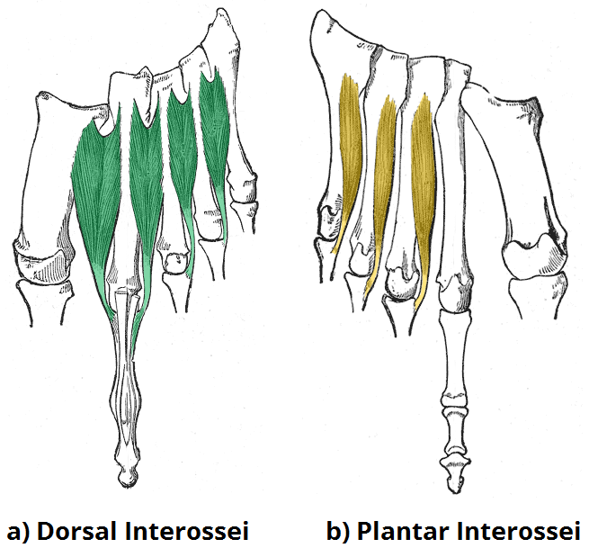
| Plantar Interossei | Fourth Layer | Originates from the medial side of metatarsals three to five. Attaches to the medial sides of the phalanges of digits three to five. | Adduct digits three to five and flex the metatarsophalangeal joints. | Lateral plantar nerve. | There are three plantar interossei, which are located between the metatarsals. Each arises from a single metatarsal. | |
| Dorsal Interossei | Fourth Layer | Originates from the sides of metatarsals one to five. The first muscle attaches to the medial side of the proximal phalanx of the second digit. The second to fourth interossei attach to the lateral sides of the proximal phalanxes of digits two to four. | Abduct digits two to four and flex the metatarsophalangeal joints. | Lateral plantar nerve. | There are four dorsal interossei, which are located between the metatarsals. Each arises from two metatarsals. |